Chromosomes are long strands of DNA found in the cells of the body. DNA contains genes that provide the code for our bodies to grow, develop and function normally. Karyotyping, or chromosome analysis, is a useful test in the diagnosis and management of fertility issues.
What are cells?
Our bodies are made up of billions of cells. There are many different kinds of cells, including blood cells, skin cells, and muscle cells. Our genetic material is contained in a structure within the cell called the nucleus. Every cell in the body, with the exception of mature red blood cell, has its own nucleus containing a complete set of genetic material.
What's inside a cell nucleus?
Inside the nucleus of the cells there are 46 rod-shaped structures called chromosomes. The chromosomes come in pairs, 23 coming from the mother and 23 coming from the father. Scientists label the chromosomes with numbers according to their size chromosome number 1 through to chromosome number 22. The 23rd pair of chromosomes are the sex chromosomes, y two X's for a female or one X and one Y chromosome for a male. The chromosomes vary in size with chromosome number 1 being the largest and chromosome number 22 the smallest of the numbered chromosomes.
What's inside a chromosome?
Each chromosome contains hundreds or thousands of genes, depending on the size of the chromosome. Genes are units of heredity and are constructed of DNA. For the most part, individual genes are too small to be seen even through a powerful microscope. Chromosomes, however, can be seen under a microscope.
What is a karyotype?
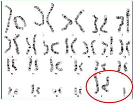
A karyotype is a photograph of all of the chromosomes of an individual cell. The scientists examining a karyotypel ine up the chromosomes lined up in pairs, so that they are in order from the largest to the smallest. A photograph is taken through a microscope, so that the longest chromosomes usually appear to be about an inch long. In reality, of course, they are so small that they are not visible to the naked eye.
How is a karyotype analysis performed?
Karyotypes are usually constructed by laboratory scientists and analysed by cytogeneticists who are trained specialists in the study of chromosomes. The cells must be grown and advanced to a specific cell stage that is optimal for analysis. The process of growing the cells, dropping them onto slides, arranging the chromosomes into a karyotype and analysis by a cytogeneticist usually takes 1-2 weeks in our laboratory.
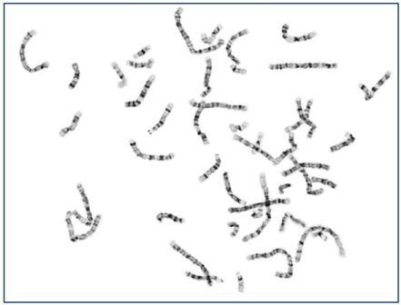
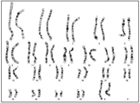
Normal female karyotype 46, XY Metaphase (pictured above)
Normal female karyotype 46, XY Karyotype (pictured above)
What can a karyotype test tell us?
Karyotype analysis determines the number of chromosomes in the cells and whether there are any pieces of chromosomal material that are missing, extra or rearranged. Any variation from the normal chromosome number and arrangement can have implications for a person's fertility and the risk for having a baby with birth defects.
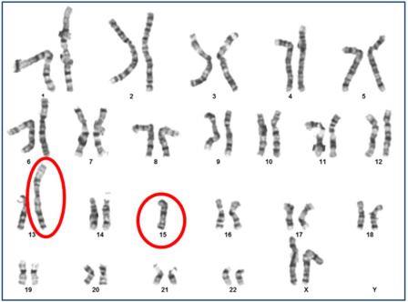
Chromosome translocation between 13/15 (pictured above)
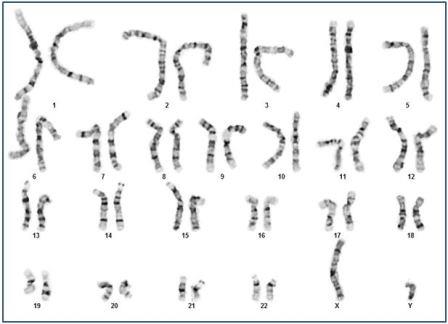
Sex Chromosome Variation, XXY (pictured above)
What can't a karyotype tell us?
Karyotype testing for both partners having difficulty conceiving or having recurrent miscarriages can provide y useful information in determining the potential cause of infertility and the most appropriate reproduction options. Knowledge of chromosomal variations can prevent the use of treatments with a high chance of failure, and can increase your chance of having a healthy baby.